
Scottish Heritage, Exceptional Breeds
Breeding with Tradition and Care for Over 20 Years.
Latest Puppy News
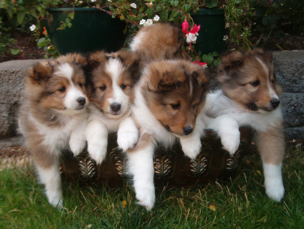
TRICOLOR / BI-BLACK SHELTIES – FALL 2025: Planned AOAC Sheltie litter which may produce Tricolor and/or Bi-Black (Black and White) Shelties. Sire: Am/Can CH Miquelon Life of Leisure “Larry” x Dam: Salt City Glamour at RavenWyn “Jo Jo”. We are currently accepting reservations with a deposit.
ROUGH COLLIES – No litters planned at the moment. Sign-up for our “Collie Pupdates” to be notified when we do plan a litter [here].
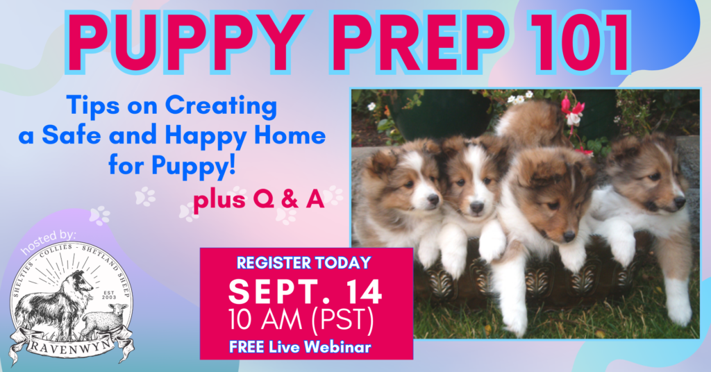
If you would like your home to be considered for a puppy and to start the application process, please complete our online Puppy Questionnaire.
Available Shetland Sheep
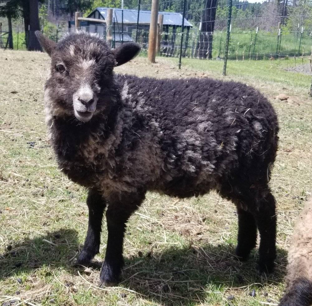
We currently have NASSA-registered rams and ewes available (possibly ewe lambs), perfect for breeding programs or enhancing your flock. For those interested in fiber arts, we also have multiple unregistered ram and ewe Wool Fiber pets that produce luxurious, soft wool ideal for spinning and crafting.
Our Shetland Sheep are an integral part of our goals toward sustainable farming. We have award-winning Shetland lines that produce fine fleece with a lot of crimp for those that enjoy spinning wool. If you are looking for sweet-tasting, Grass-fed lamb in Oregon, Washington or Idaho, we can accommodate that as well. Contact us for details on available Shetland Sheep
Preserving Scottish Heritage at RavenWyn
Nestled on a serene homestead just 30 minutes east of Clackamas, Oregon, our family is dedicated to raising and nurturing some of the most beloved Scottish breeds. Surrounded by the beauty of the Pacific Northwest, our home provides an ideal environment for our dogs and sheep to thrive. Read More…